CFD and FEM-based Aneurysm Research latest developments and a course on Big Data analysis at the MeDiTATe Summer School in Tinos
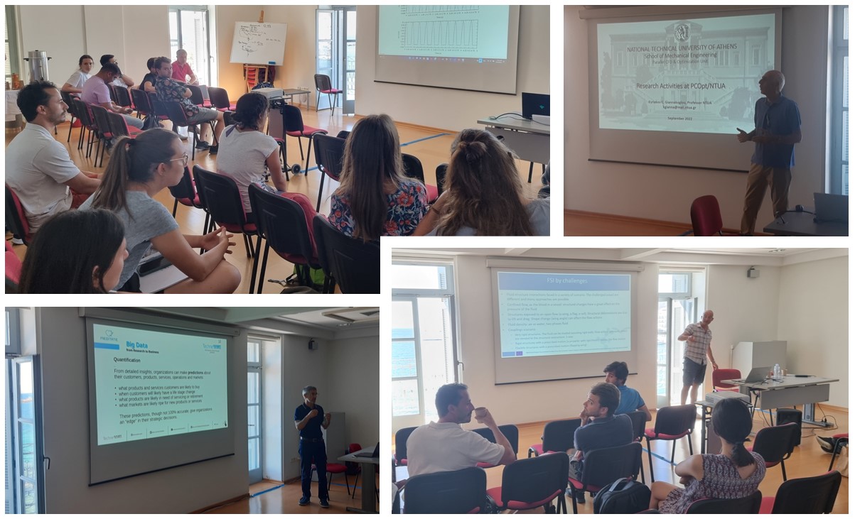
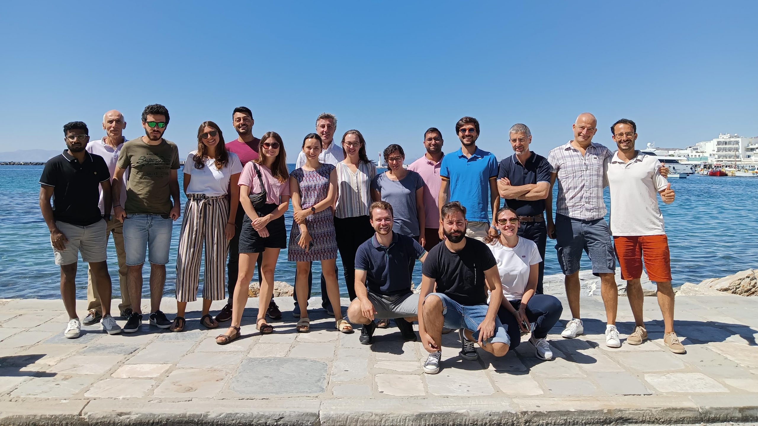
The National Technical University of Athens hosted the MeDiTATe project Summer School on CFD/FEM-based aneurysm research and big data analysis. The event took place from 12th to 16th September 2022 on the Island of Tinos, in Greece.
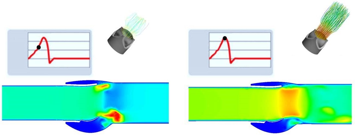
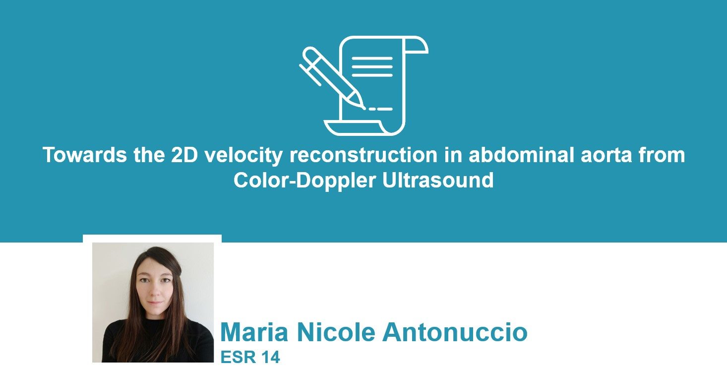
The Early Stage Researchers of the MeDiTATe project had the opportunity to follow several courses and conferences on CFD, FEM, Fluid-Structure Interactions, big data and their application to hemodynamics and aneurysms studies.
The summer school was also an opportunity to strengthen the collaboration among the Early Stage Researchers and make contact with other members and partners of the consortium.